Biomembranes enclose living cells and are found in the intracellular organelles. Their bilayer structures (lipid bilayers) protect the cell interior from external influences [2] and enable biochemical reactions to take place in the organelles.
Membrane lipids
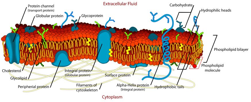
Biomembranes (Fig. 1) consist mainly of phospholipids, sphingolipids and cholesterol – the membrane lipids. The proportionally largest group of phospholipids forms amphiphilic molecules (ancient Greek: amphí = on both sides, phílos = loving), which contain hydrophilic (water-loving) "heads" and two hydrophobic (fat-loving) "tails". The heads consist of phosphate groups to which choline, ethanolamine, glycerol, serine or inositol are attached, and project either from the inner layer of the bilayer into the cell interior or from the outer layer into the cell surface. The hydrophobic fatty acid residues of the tails are directed towards the interface of the two adjacent layers of the bilayer.
The cell surface is dominated by phosphatidylcholine and sphingomyelin, which belongs to the sphingolipids. Phosphatidylserine, phosphatidylethanolamine and phosphatidylinositol are found on the inside of the bilayer. [2, 3, 4, 5]

Fig. 1: Structure of the cell membrane [6].
Change of composition
The inner and outer layers of the bilayers do not only generally differ in their compositions. The compositions can also change locally for a short time in response to external stimuli, such as injuries. For example, phospholipids of the inner layer change into the outer layer. In this way, the cell can react to stimuli from the environment. [7, 8]
Membrane proteins
The exchange of substances and communication between the cells and their environment are mainly controlled by proteins that are incorporated into the membranes. Transmembrane proteins have contact with the cytosol, the medium inside the cells, and with the extracellular space. [9] They form pores, channels and carriers. The cytosol, for example, contains low concentrations of calcium ions, whereas their concentrations in the extracellular space are comparatively much higher. Changes in concentration gradients trigger intra- and extracellular processes. [10]
On the inside and outside of the bilayers are other proteins that are coupled to lipids or to transmembrane proteins. [7, 9]
Necrosis
Cells die by necrosis or apoptosis. Necrosis is a pathogenic and unregulated process. Injuries cause the biomembrane to lose its protective function. Ions from the extracellular space can flow in unhindered, and conversely, cell components reach the outside. There, they trigger an inflammatory reaction and activate the immune defence. The breakdown of the osmotic balance causes the cell to expand and rupture. The released cell components are eliminated by phagocytes ("scavenger cells"). [11]
Apoptosis
Apoptosis proceeds according to a regulated programme. Caspases, a group of enzymes activated during apoptosis, are responsible for this. Phosphatidylserine concentrates on the outer surface of the cell and attracts phagocytes. [4, 12] The cell begins to shrink (apoptotic volume decrease). The cell organelles are enclosed in vesicles (apoptotic corpuscles). They are still surrounded by an intact biomembrane. Cell components do not enter the extracellular space and no inflammatory reaction is triggered.
Current research assumes that the phagocytes have a special receptor for phosphatidylserine. This enables them to quickly identify and remove the cells to be removed. [8, 9, 11, 12]
Conclusion
The concentration of phosphatidylserine on the outside of biomembranes prevents inflammatory reactions and ensures the effective removal of dead cells by phagocytes. [13] Studies show that during apoptosis, messenger substances that trigger inflammation, such as cytokines, are suppressed and TGF-ß (transforming growth factor) as well as prostaglandin E2 are generated. [14]
Literature
- P. Fritsch, Dermatologie und Venerologie: Lehrbuch und Atlas, Springer-Verlag, Berlin 2013, ISBN 978-3662217719
- R. Winter, Chemie in unserer Zeit 1990 (2), 71-81
- I. Bernhardt, Biol. Unserer Zeit 2007 (37), 310-319
- B. Jacobi, S. Partovi, Basics Molekulare Zellbiologie, Elsevier Urban & Fischer Verlag, München 2011, ISBN 978-3-437-42686-5
- M. Ebara, Chonnam Medical Journal 2019 (55), 1-7
- LadyofHats, Public domain, via Wikimedia Commons, "Zellmembran"
- P.J. Sims, T. Wiedmer, Thromb Haemost 2001 (86), 266-275
- E. M. Brever, P.L. Williamson, Physiological Reviews 2016 (96), 605-645
- R. Lüllmann-Rauch, F. Paulsen, Taschenlehrbuch der Histologie, Georg Thieme Verlag KG, Stuttgart 2012, ISBN 978-3-13-129244-5
- B. Alberts et al., Molekularbiologie der Zelle, VCH Verlagsgesellschaft mbH, Weinheim 1990, ISBN 978-3527279838
- C. D. Bortner, J. A. Cidlowski, Frontiers in Cell and Developmental Biology 2020 (8), 1-16
- V. A. Fadok et al., Cell Death and Differentation 1998 (5), 551-562
- R. B. Birge et al., Cell Death and Differentation 2016 (23), 962-978
- P. M. Henson, D. L. Bratton, The Journal of Clinical Investigation 2013 (123), 2773-2774
Anne Schieferecke |


