The external skin layer, called epidermis, is a dynamic system of continuously proliferating and differentiating activity. It mainly consists of keratinocytes, melanocytes and immunocompetent cells (Langerhans cells). Structure and metabolism of the epidermis have two essential functions: to protect the skin from external influences and to maintain the hydration and the osmotic balance of the inner tissues. Both functions are accomplished by the stratum corneum which is the outermost of the 5 epidermis layers.
Pluripotent native cells are formed in the basement membrane. They differentiate into keratinocytes within the following layers and secrete the dermal protein keratin as well as different lipids from keratohyalin and lipid granules.
During their ripening process and their migration towards the skin surface, the keratinocytes modify their metabolism and their morphology. Flat keratinocytes without nucleus form the "bricks" of the protective wall and are now called corneocytes. The stratum corneum consists of about 60 % structural proteins, 20 % water and 20 % lipids. Intracellular lipid lamellae consist of ceramides, cholesterol, phospholipids and long-chained free fatty acids and form the „mortar" in this barrier. Ceramides tightly link to the horny shell of keratinocytes and hence stabilize the lipophilic hydration barrier. Within the transitional zone, there are intense enzymatic activities which lead to the release of lipids out of the granules and to their chemical modification. Just to mention an example: the barrier strengthening gamma linolenic acid is formed from the essential linoleic acid. The barrier function of the stratum corneum depends on the specific lipid composition. Disorders of the barrier function result in skin diseases as i.e. atopic dermatitis, psoriasis, acne, dry and sensitive skin or premature skin aging processes.
Antimicrobial peptides
Specific intracellular lipids, sebum, the natural moisturizing factor (NMF), organic acids and inorganic ions control the moisturizing capacity of the stratum corneum. While migrating from the stratum basale to the stratum corneum, the corneodesmosome links between the keratinocytes are degraded by enzymes (filaggrin and peptidylarginine deiminase) with the effect that the densely packed keratinozyte layers are loosened and corneozytes are chipped off in an orderly desquamation process. The peptides and amino acids which result from the corneodesmosomes and filaggrin degradation form the natural moisturizing factor (NMF) of the skin barrier. Both ripening process of the keratinocytes and function of the stratum corneum are primarily influenced by the moisture content of the epidermis [Farage MA et al., Textbook of Aging Skin, Springer-Verlag 2010]. Two important antimicrobial peptide families (AMPs) are expressed within the stratum corneum, i.e. ß-defensine (hBD1,2,3 and 4), which are cathelin-related antimicrobial peptides (CRAMP) and the cathelicidin-carboxy-terminal fragment (LL-37). The peptides hBD2 and LL-37 are secreted from lamellar bodies together with the lipids. Along with the keratinocytes, they are the natural antibiotics of the skin and form a solid defense against microbes [Oren A et al., Exp Mol Pathol 2003; 74: 4271-78; Schauber J et al. J Invest Dermatol 2007; 127: 510-12]. In case of inflammatory skin diseases and wounds the AMP concentration will significantly increase.
Rosacea is an inflammatory skin condition characterized by papulopustular erythema and teleangiectasia. Yamasaki K et al. [Nat Med. 2007; 13(8): 904-6] could prove that rosacea involves high concentrations of modified cathelicidins. These cathelicidin peptides are associated with a significant increase of tryptic enzymes (serine proteases) in the stratum corneum and initiate the well-known rosacea skin inflammations. The genetic cause for the formation of rosacea is a deletion in the cathelicidin coding gene CAMP. In case of acute barrier disorders, the hBD2 and CRAMP concentration increases and a cytokine cascade is released. At the same time, lipids are secreted from the granules of the stratum corneum in order to repair the damaged barrier. Hence, an intact stratum corneum is a permeability or hydration barrier and also acts as an inviolable obstruction for microbes. [Park KY et al. J Korean Med Sci 2010; 25: 766-771; Elias PM et al J Invest Dermatol 2008; 128: 1067- 70; Elias PM et al Am J Contact Dermat 1999; 10:119-26].
While LL-37 acts as an antimicrobial peptide within the epidermis at the one hand, it controls the fluidity of the lipid bilayer of the skin barrier by modulating the lipid synthesis on the other hand. Hong SP et al. [J Invest Dermatol 2008; 128:2880-2887] proved that a sub-erythemal UV radiation releases antimicrobial peptides in the epidermis, repairs barrier disorders and improves the resistance to microbes. Henkel A et al. [Sci Pharm. 2009; 77: 238] showed via "target-fishing strategy" that boswellia acids (frankincense acids) specifically link onto LL-37 and thus improve and modify the anti-inflammatory effect of AMP. In addition to that, LL-37 induces the release of barrier strengthening lipids.
There are other topically applied phyto preparations as eg betulins or betulinic acids which can stimulate the expression of AMP in the skin and the mucous membrane. That implies that herbal extracts offer new mechanisms of immunostimulation and barrier strengthening processes.
Stressed skin - stress proteins and heat shock proteins (HSP)
Heat shock proteins were first observed by Ritossa [Drosophila Experienta 1962; 18: 571-3] who discovered them in fruit flies of the species drosophila melanogaster after temperature changes. In the late 1970s, it turned out that the heat shock reaction is not a specific feature of certain cells or organisms but a universal protective mechanism of the cells.
Stress proteins and heat shock proteins form a specific group of proteins and occur in pro- and eucaryotic cells. Depending on their molecular size and function, they are divided into five different families. HSPs are induced by various environmental influences that endanger the cell life as eg heat, cold, hypoxemia, oxidative and nitrosative stress, inhibitors of the mitochondrial energy metabolism and also by harmful substances.
Heat shock proteins also occur in "unstressed" body cells, however in low concentrations only. There they act as chaperones (protein folders) which are responsible for the proper function of the cell proteins at the right time and at the proper position. Damaged proteins are repaired and folded into their active form. The HSPs control proliferation, differentiation and apoptosis processes in the cells. The HSPs "accompany" the proteins from one cell compartment into another and signalize damaged peptides to the immune system.
Lindquist S [Annu Rev Biochem 1986; 55: 1151-91] discovered the induction of thermal tolerance as the main function of the heat shock proteins, an inducible phenomenon which means that after the once experienced heat stress, the cells become resistant to an otherwise lethal heat stress. Maytin E et al. [J. Invest. Dermatol 1990; 95: 635-42; J. Biol Chem. 1992; 267: 23189-96; J. Invest Dermatol 1995; 104 (4): 448-55] proved that heat stress in human keratinocytes will not only lead to thermal tolerance and show an improved heat shock protein synthesis but also involve insensitivity to other stresses and strains as for instance those caused by heavy metals and hypoxemia. Surprisingly, it also leads to a resistance against UVB induced damages. Furthermore, it was observed that after the hyperthermia treatment, additional HSP 72 is expressed on the cell surfaces of tumor cells which results in an increased immunogenicity. The heat shock proteins use thermal stress to adjust the natural defense activities against tumor cells.
Radiofrequency, laser or IPL (Intense Pulsed Light) treatments of the skin can simulate thermal stress and thus activate the release of antimicrobial peptides and heat shock proteins. The induction of heat shock proteins is one of the manifold molecular-biological effects of the hyperthermal treatment. Schroeder P et al. [J. Invest. Dermatology 2008; 128, 2491-2497] described though, that the infrared radiation (IRA; 760-1.440 nm) releases the collagen-degrading metalloproteinase-1 (MMP-1) in the dermis but not epidermis, and leads to a significant reduction of antioxidants in the skin. Topical antioxidants as for instance the vitamins A, C, E, or epigallocatechingallate could avoid the collagen-damaging effects of MMP-1 on the skin. Boswellic acids (acetyl-keto-β-boswellic acid) also are specific inhibitors of the matrix-metalloproteinases.
Inflammatory skin diseases and skin aging
Structural and molecular-biological changes occur within the skin over the years. The stratum corneum becomes thicker and the hydration barrier is damaged by cornification and pigment disorders. Atopic dermatitis, actinic keratosis, psoriasis, rosacea and other skin diseases are characterized by barrier damages and chronic inflammations. The heat shock protein synthesis is reduced and the repair mechanisms of the skin are disordered. Damaged proteins and peptides are no longer repaired or disposed of. The controlled apoptosis of the keratinocytes is impaired and the immune defense is weakened. Simultaneously to these processes, inflammatory enzymes as e.g. the matrix-metalloproteinases are formed. Skin aging and skin inflammations hence are characterized by the fact that the repair mechanisms of the skin are disordered and enfeebled and that additional inflammation mediators are released. That is the reason why dermatocosmetic anti-aging treatments of the aging and inflamed skin have to take account of these molecular-biological processes.
Jabs HU [Ästhetische Dermatologie 2009; 4: 28-33] described an improved skin structure after an innovative anti-aging treatment based on intense pulsed light (IPL) and boswellia nanoparticles. Frankincense contains anti-inflammatory boswellic acids which can be additionally activated by infrared light. Henkel et al. (2009) could prove that boswellic acids specifically bind to the antimicrobial peptide LL-37 of the skin and thus control the anti-inflammatory features. The disordered lipid synthesis of the skin barrier is improved with the help of LL-37 and ceramides are released from the lipid granules. Boswellic acids (acetyl-keto-β-boswellic acid) specifically inhibit the enzymes of the inflammatory cascade as e.g. inflammatory 5-lipoxygenases and collagen-degrading matrix-metalloproteinases. The IPL flashes exert thermal strain on the skin, a fact that stimulates the HSP synthesis. The innovative anti-aging treatment with IPL activated boswellia nanoparticles positively influences skin aging processes in two different ways: heat shock proteins and antimicrobial peptides are stimulated into protective factors on the one hand, on the other hand, inflammatory processes and collagen disorders are reduced and barrier disorders repaired.
A disadvantage of the IPL treatment is that the energy of the flash has to be reduced for tanned skin. That is why a procedure was searched that stimulates the heat shock proteins and antimicrobial peptides and activates the boswellic acids also on tanned skin.
Radiofrequency & boswellia treatment - an innovative anti-aging strategy
A radiofrequency treatment with filtered 2.3 MHz waves permits to specifically warm up the skin and stimulate the HSPs and AMPs. The electromagnetic radiation additionally activates the boswellic acids.
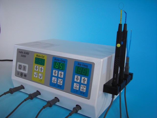
radioSURG® 2200 (Meyer-Haake GmbH, Wehrheim) is a radio frequency device designed for radiosurgery, and its frequency of 2.2 MHz permits to generate different modulated and non-modulated high frequency currents. The device has a filtered wave and various coagulation settings and is used for different types of tissue-conserving radiosurgery.
In combination with a specific cone electrode and filtered non-modulated high frequency current, the device can be used for the specific radiofrequency therapy of the skin. The energy of the radio waves arrives at the dermis and causes a shrinking of the stretched collagen and elastin fibers. This has a smoothing effect on the wrinkles. In the epidermis, it releases heat shock proteins and antimicrobial peptides.

Fig. 1: radioSURG® 2200 radio frequency device (Meyer-Haake, Wehrheim)
Boswellia nanoparticles
Frankincense (Boswellia carteri, Boswellia sacra) is a resin gained by cutting into the bark of desert trees of the boswellia species. Main growing area of the boswellia trees is the Middle East and primarily Oman, Yemen, Somalia and India. The leaking resin hardens in contact with atmospheric oxygen; it is painstakingly hand-harvested with a special scraping knife and traded in frankincense bazars.
Frankincense extracts have anti-inflammatory and anti-allergenic features and also show anti-tumor activity. They are very effective against inflammatory skin diseases as e.g. actinic keratosis, psoriasis, atopic dermatitis and acne.
According to current knowledge, the boswellic acids are the pharmacologically effective components among the ingredients of the frankincense resins. Sashwati et al. [DNA and Cell Biology 2005;24 (4): 244-255] proved the anti-inflammatory and collagen protecting mechanism of acetyl-keto-boswellic acids. They inhibit the collagen degrading matrix-metalloproteinases as well as the inflammatory 5-lipoxygenases in the skin.
A standardized frankincense extract with a minimum content of about 30 % acetyl-keto-ß-boswellic acids has been isolated to treat inflammatory skin diseases and skin aging conditions. The resinous and extremely sticky active agent concentrate has been encapsulated into nanoparticles and thus made available for the penetration into the skin (KOKO GmbH & Co. KG, Leichlingen).
Nanoparticles are spherical bodies of the size of 60-100 nm with a shell consisting of the natural phospholipid phosphatidylcholine of the skin. By means of a specific galenic procedure, lipophilic substances can be encapsulated into nanoparticles where they evolve into a watery dispersion. After the nanoparticles are applied on the skin, the PC shell assimilates with the lipid bilayer of the stratum corneum and the content of the nanoparticles is transported into the deeper layers of the skin. Certain active agents can only pass through the skin barrier with the help of this specific transport mechanism.
Boswellia nanoparticles can be integrated into a DMS cream (Derma Membrane Structure, KOKO, Leichlingen). The result is a barrier strengthening skin care cream which is rich in active agents and used to treat inflammatory and proliferative skin diseases.
Boswellia nanoparticles can be mixed with a specific cosmetic gel base which is free of preservatives (KOKO, Leichlingen) in a 1:1 ratio (gel:active agent). This boswellia gel was applied for the radio frequency treatments.
Treatment sequence
After the cleansing of the skin, the boswellia gel is applied with a brush onto the specific skin area.
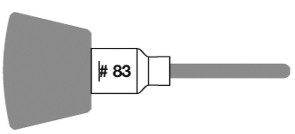
The hand piece is equipped with a specific RF cone electrode. The electrode helps to spread the radio waves evenly onto the desired area and penetrate them into the deeper skin layers together with the boswellic acids.
For the treatment of facial, neck and décolleté areas, the neutral electrode is placed underneath the shoulder of the patient. Attention should be paid on the fact that the neutral electrode is entirely covered by the patient. A direct skin contact is not required as the electrode serves as antenna. Only high frequency devices in the Mega Hertz range have a sophisticated radio waves leakage so that a skin contact is not required. There will be no burns, which are feared in connection with kilohertz devices. The radioSURG® 2200 is set to MONO CUT (no coagulation rate) and then adjusted to 6 to 10 Watt (no local anesthetic) or 14 to 18 Watt (with local anesthetic).
 Fig. 2: Cone electrode for the radio wave & boswellia treatment
The electrode is gently pressed onto the skin area in order to avoid gaps between electrode and skin. Only then, the device is turned on via foot or hand switch while the electrode is moved in circling movements over the specific skin area. It is required to slightly press the electrode onto the skin in order to minimize the light heat and pain sensation that is felt when the electrode is only lightly applied. The more intense sensation of pain felt with a lightly applied electrode is due to the potential spark discharge in the gap between skin and electrode. Yet, this is a physical law and applies to all radio frequency units. The spark discharge is invisible though, but it is felt like a light stitch. It is important to constantly move the electrode.
In general, a range of 10 to 14 Watt is well tolerated on the cheeks. However, the patients prefer lower settings around the eyes and on décolleté and upper lip. The setting depends on the personal experience of pain of the individual patient and the thickness of the skin. If an anesthetic is used, the individual settings may be eventually increased up to 20 Watt.
It is suggested to do a tolerance test before every treatment. If the skin turns lightly red around the décolleté, the setting is perfect for the more sensitive parts of the skin.
Cooling gels or traditional ultrasound gels should not be used as the perfume and preserving components contained may have an allergenic and sensitizing effect on the skin that could be intensified by the radio waves. The skin areas were systematically treated with circling movements. Extremely deep wrinkles (glabella, mimic and nasolabial wrinkles) were treated by following the course of the wrinkle with small spherical electrodes or thick needle electrodes. Due to the small surface of spherical and needle electrodes, more heat is generated in this case and that is the reason why a lower setting has to be chosen. The intense wrinkle treatment may lead to the formation of scabs in isolated cases. It should never be attempted to remove the scab as scars might form otherwise.
The treatment of the face, neck and the décolleté takes about 30 minutes. After the treatment, the skin is lightly red and the wrinkles are padded. Immediately after the treatment, there is a noticeable and visible skin condition to observe. However, it is recommended to inform the patients on the fact that this immediate effect will disappear after about 24 hours. The proper buildup of the collagen and elastin structures will occur in the deeper layers of the skin and be visible on the epidermis about 8 to 10 days after the treatment. This condition will even improve over a period of 3 months.
The following cream formulation is handed out to the patients for the skin care at home:
dermaviduals® High Classic Plus membrane cream (44 ml, KOKO, Leichlingen) is mixed with
-
2 ml Boswellia nanoparticles
-
2 ml liposome concentrate plus with acelaic acid
-
2 ml nanoparticular butcher's broom extract
The radio wave & boswellia method can be combined with any other dermatocosmetic treatment. With the proper use of device and procedure, no side effects have been observed so far. Younger patients frequently need one session only. Elderly patients suffering from skin diseases are recommended to undergo 6 treatments with a 1 to 2 weeks' interval. Experience has shown that the effects will last for about 9 to 15 months.
Results and discussion
Objective of this study was to find out whether the treatment with radio waves and boswellia nanoparticles could achieve the same results as the treatment with IPL activated boswellia nanoparticles. It could be proved that the innovative anti-aging treatment based on radio waves and boswellia nanoparticles is very effective and that the results could well be compared with those achieved by the IPL-boswellia procedure. In contrast to the IPL boswellia method, however, the radio frequency treatment could also be applied on tanned skin. The radio wave triggered heat stimulus simulates burns within the skin which lead to an intensified synthesis of antimicrobial peptides and heat shock proteins in the epidermis. The effective anti-aging treatment here actually consists of a stimulation of the natural repair mechanism of the skin. In addition to that, the radio waves tighten the collagen and elasticin fibers and stimulate the collagen synthesis.
Boswellia nanoparticles support the repair of the skin barrier by linking with the antimicrobial peptide LL-37, and by inhibiting the inflammatory 5-lipoxygenases and the collagen degrading matrix-metalloproteinases. Phosphatidylcholine which is released from the nanoparticle shells is a natural phospholipid of the skin which stabilizes the membranes in the epidermis and stimulates the ceramide synthesis. The energy of the electromagnetic radio waves additionally stimulates and activates the triterpenes of the boswellic acids.
Due to the induction of antimicrobial peptides and heat shock proteins, the radio wave boswellia treatment has excellent preventive effects against UV damages of the skin. It can intensify the effect of sun protection creams by inducing HSPs as protective factors as well as by the fact that boswellic acids neutralize free radicals that form after UV absorption of the light protection filters.
Based on the biochemical mode of action, it can be assumed that the method can also be used as preoperative treatment for the surgery of proliferative skin diseases as e.g. melanomas.
In conclusion, it can be maintained that the radio wave & boswellia method is an innovative and highly effective cosmetic procedure to rejuvenate the skin and to treat inflammatory skin diseases. Radio wave & boswellia reverse the skin aging mechanisms and stimulate the natural repair mechanisms of the skin with natural herbal extracts.
Dr. Hans-Ulrich Jabs | 
